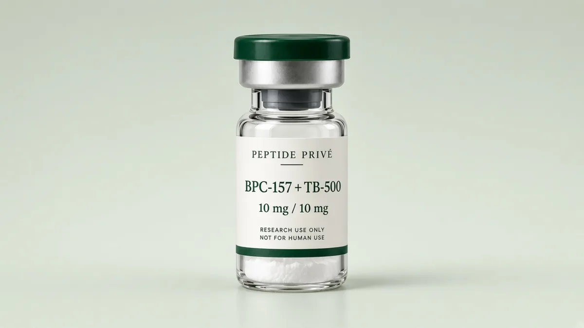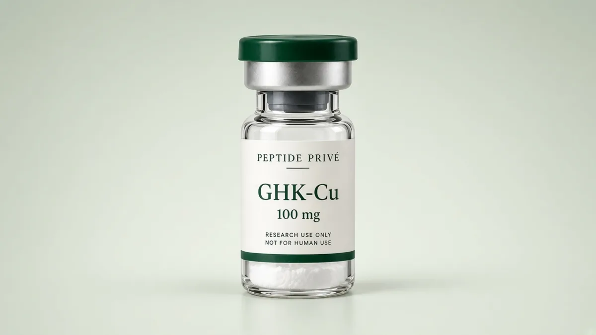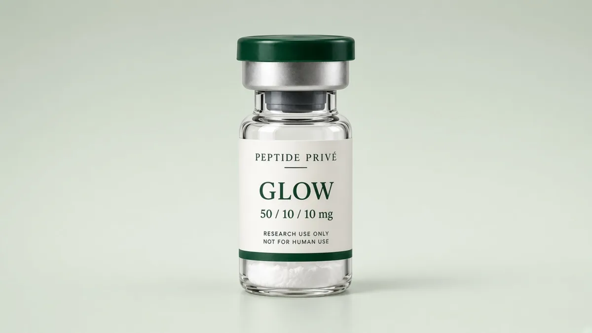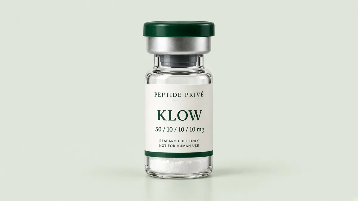
A multi-peptide blend places two or more distinct peptides in a single vial at fixed amounts. Reading its label correctly is the first step in keeping your lab records accurate, and it shapes how the vial should be analyzed and documented.
What a Blend Is
A blend is a co-packaged combination of separate compounds, not a new molecule. Each peptide keeps its own sequence, Chemical Abstracts Service (CAS) number and molar mass. The label simply tells you how much of each one the vial contains.
The components are not chemically bonded to each other. In the dry powder they sit side by side; once dissolved, each one behaves according to its own chemistry. One may be more sensitive to light, another to pH, a third to surface adsorption. A blend's storage conditions have to suit its most sensitive component.
How the mg Amounts Map to Each Component
The amounts on a blend label are listed in the same order as the component names. Our GLOW blend is a clear example.
| Component | Amount per vial |
|---|---|
| GHK-Cu | 50 mg |
| BPC-157 | 10 mg |
| TB-500 | 10 mg |
| Total | 70 mg |
The same logic applies to two-part blends. BPC-157 + TB-500 contains 10 mg of each peptide.
Mass Ratio Is Not Molar Ratio
Labels are written in milligrams, but molecules are counted in moles. Because peptides differ widely in size, a mass ratio can hide a very different molecular ratio.
Take GHK-Cu (molar mass 401.91 g/mol) and BPC-157 (1419.55 g/mol) in GLOW:
| Component | Mass | Molar mass | Amount of substance |
|---|---|---|---|
| GHK-Cu | 50 mg | 401.91 g/mol | about 124 µmol |
| BPC-157 | 10 mg | 1419.55 g/mol | about 7.0 µmol |
The mass ratio is 5 to 1, but the molar ratio is roughly 18 to 1. If your experiment is designed around molar concentrations, convert each component separately rather than scaling from the label.
These figures use the nominal label mass. As explained below, the weighed powder also contains counter-ions and water, so the true peptide amount of each component is somewhat lower than the label mass.
One Identity Check per Component
A single purity figure describes the whole vial, but identity must be confirmed for every peptide inside it. On the analytical report, look for a separate mass spectrometry (MS) result for each component, matching its expected molar mass. See How to Read a Peptide Analytical Report for a section-by-section walkthrough.
Why blends are harder to analyze
- More peaks, more overlap. In reversed-phase HPLC each component gives its own main peak, and each brings its own family of related impurities. Those families can overlap with another component's main peak.
- "Purity" needs a definition. For a blend, a report might give the purity of each component peak or one combined figure. Ask which it is. A combined figure cannot tell you whether one component is clean and another is not.
- LC-MS helps. Coupling chromatography with high-resolution mass spectrometry lets an analyst identify and quantify species even when they co-elute, because they are separated by mass as well as by retention time (Zeng et al. 2015). In ESI spectra each component also produces its own set of charge-state peaks (Fenn et al. 1989), so a deconvoluted mass for each component is the clearest identity record.
- Small molecules behave differently. A tripeptide-copper complex such as GHK-Cu is far smaller than a 15-residue peptide. It elutes and ionizes differently, and the copper changes how it appears in the mass spectrum. Analysts often check such components with a method tailored to them.
Counter-Ions and Water in a Blend
Peptides purified by reversed-phase HPLC are typically isolated as salts, most often trifluoroacetate, and lyophilized powders also hold residual water (Sikora et al. 2018). In a single-peptide vial, that only affects one compound. In a blend, each component may carry a different counter-ion load. The label mass of each component is the intended amount of that material, not a measured net peptide content.
Counter-ions matter in cell work too. Trifluoroacetate at low concentrations has been shown to reduce proliferation in osteoblast and chondrocyte cultures, which led the authors to recommend converting peptides to a hydrochloride or comparable salt before biological assays (Cornish et al. 1999). If your cell model is sensitive, record the salt form of each component.
Label Amounts and Stock-Solution Concentration
When a blend is dissolved in a laboratory solvent, every component is diluted by the same volume. Concentration per component equals its mg amount divided by the total solvent volume. For documentation, if a GLOW vial is dissolved in 5 mL of laboratory solvent:
- GHK-Cu: 50 mg / 5 mL = 10 mg/mL
- BPC-157: 10 mg / 5 mL = 2 mg/mL
- TB-500: 10 mg / 5 mL = 2 mg/mL
The ratio between components never changes, only the concentration of each. This is identity and accounting math for the lab notebook, not a use protocol.
Recording a Blend in Your Notebook
| Field | Why it matters |
|---|---|
| Blend name and lot number | Links the vial to its report |
| Each component and its label mass | The basis for every concentration you calculate |
| Molar mass of each component | Needed for molar conversions |
| Solvent and volume | Defines the stock concentration |
| Date prepared and storage | Lets you track age of the solution |
Storage
Store sealed blend vials cold, dry and away from light. After a solution is prepared for laboratory work, refrigerate it and record the date, volume and lot number on the label. Because a solution stores several peptides together, follow the most conservative storage requirement among the components. See Storing Lyophilized Peptides in the Lab.
Every Peptide Privé vial is batch-numbered. Every Peptide Privé batch is tested by an independent lab. Shop all peptides or contact our team with any labelling questions.
Sources
- Zeng K et al. Liquid Chromatography-High Resolution Mass Spectrometry for Peptide Drug Quality Control. AAPS J. 2015. PubMed 25716148: https://pubmed.ncbi.nlm.nih.gov/25716148/
- Fenn JB et al. Electrospray ionization for mass spectrometry of large biomolecules. Science. 1989. PubMed 2675315: https://pubmed.ncbi.nlm.nih.gov/2675315/
- Sikora K et al. Counter-ion effect on antistaphylococcal activity and cytotoxicity of selected antimicrobial peptides. Amino Acids. 2018. PubMed 29307075: https://pubmed.ncbi.nlm.nih.gov/29307075/
- Cornish J et al. Trifluoroacetate, a contaminant in purified proteins, inhibits proliferation of osteoblasts and chondrocytes. Am J Physiol. 1999. PubMed 10567002: https://pubmed.ncbi.nlm.nih.gov/10567002/
- PubChem, BPC-157 (CID 9941957): https://pubchem.ncbi.nlm.nih.gov/compound/9941957
- GHK-Cu molar mass as listed in our GHK-Cu compound profile.
For in-vitro laboratory research only. Not for human or animal use.